Starviewer is an Open-source Free Medical Imaging Software for Researchers and Radiologists
Explore Advanced Medical Imaging with Starviewer: The Ultimate Open-Source DICOM Viewer

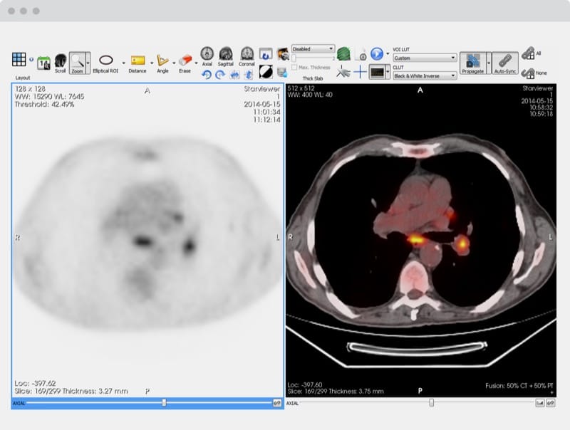
Table of Content
Starviewer is an open-source medical imaging software designed for advanced DICOM image viewing and analysis. It offers a comprehensive set of tools that enable healthcare professionals to visualize, manipulate, and analyze medical images effectively.
Starviewer is cross-platform, making it accessible on various operating systems, and it is particularly suitable for use in clinical environments, research, and education.
The community edition is totally free to download and use.
Features
- DICOM Compatibility: Fully compliant with DICOM standards, ensuring seamless integration with medical imaging systems.
- Advanced Image Viewing: Provides 2D, 3D, and multiplanar reconstructions (MPR) for detailed image analysis.
- Cross-Platform Support: Available on multiple operating systems, including Windows, Linux, and macOS.
- Image Manipulation Tools: Includes tools for zooming, rotating, and adjusting contrast, brightness, and color maps.
- ROI (Region of Interest) Tools: Allows for the precise selection and analysis of specific regions within an image.
- Annotation and Measurement: Supports adding annotations and measuring distances, areas, and volumes within images.
- Plugin Architecture: Extensible through plugins, enabling the addition of custom features and tools.
- Multi-Language Support: Offers a user interface available in multiple languages, increasing accessibility for international users.
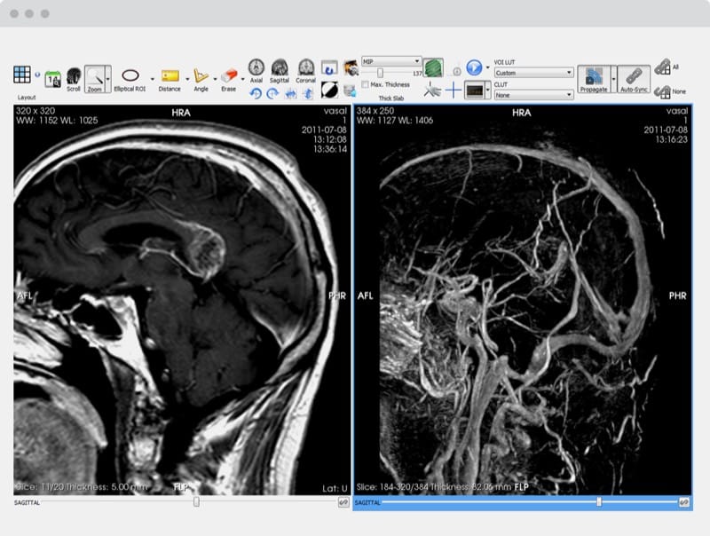
- 3D Volume Rendering: Capable of generating 3D visualizations from DICOM datasets for comprehensive image analysis.
- Export and Reporting: Allows for exporting images and generating reports, facilitating communication and documentation.
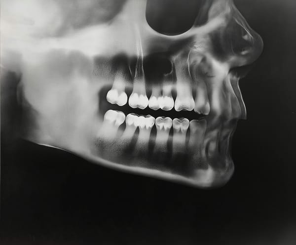
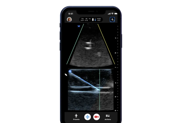
- Multi-Modality Support: Handles various imaging modalities, including CT, MRI, PET, and ultrasound, allowing for versatile medical image analysis.
- Multi-Planar Reconstruction (MPR): Enables viewing of images in multiple planes (axial, coronal, sagittal), providing a comprehensive perspective on the scanned area.
- 3D Surface Rendering: Supports creating 3D surface models from medical images, useful for pre-surgical planning and educational purposes.
- Fusion Imaging: Allows for the overlay and fusion of images from different modalities (e.g., PET-CT) to provide combined anatomical and functional information.
- Interactive Volume Rendering: Facilitates interactive exploration of 3D datasets, enabling users to adjust rendering parameters on-the-fly.
- Time Series Analysis: Supports viewing and analysis of time-series data, such as cardiac and functional MRI sequences.
- Automated Segmentation Tools: Includes tools for automatic segmentation of structures within images, aiding in faster and more accurate analysis.
- Scripting and Automation: Provides support for scripting to automate repetitive tasks and enhance workflow efficiency.
- Customizable Interface: Allows users to customize the layout and functionality of the interface to suit their specific needs.
- Integration with PACS: Seamlessly integrates with PACS systems for easy retrieval, storage, and sharing of medical images within a networked environment.
- Multi-User Collaboration: Enables multiple users to collaborate on image analysis, sharing views and annotations in real-time.
- Security and Data Privacy: Incorporates robust security features, including user authentication and data encryption, ensuring compliance with healthcare data protection standards.
Platforms
- Windows
- macOS
- Linux
Resources & Downloads
GitHub - starviewer-medical/starviewer: Starviewer, a cross-platform open source medical imaging software
Starviewer, a cross-platform open source medical imaging software - starviewer-medical/starviewer
 starviewer-medical
Starviewer
Starviewer is an open cross-platform medical imaging software fully compliant with the DICOM standard for image communication and image file formats. It integrates state-of-the-art image visualization and assessment tools needed in medical image diagnosis, but also advanced reconstruction techniques, 3D navigation tools and image fusion support. Its modular design allows the addition of new functional modules and it may be integrated with any healthcare information system (PACS/HIS/RIS).