OpenREM is a robust, cost-free, and open-source software application specifically designed for the critical task of patient dose monitoring.
This sophisticated tool provides a comprehensive suite of functionalities for efficient data gathering and optimization of radiation exposure, thereby supporting the goal of minimizing patient risk while ensuring effective medical imaging.
Key Features
One of the key features of OpenREM is its capability to seamlessly import data from a wide range of imaging modalities, a versatility that makes it a valuable asset in diverse clinical settings. Once the data is imported, OpenREM presents summary exposure data in an easily comprehensible format, enabling healthcare providers to quickly assess the level of radiation exposure.
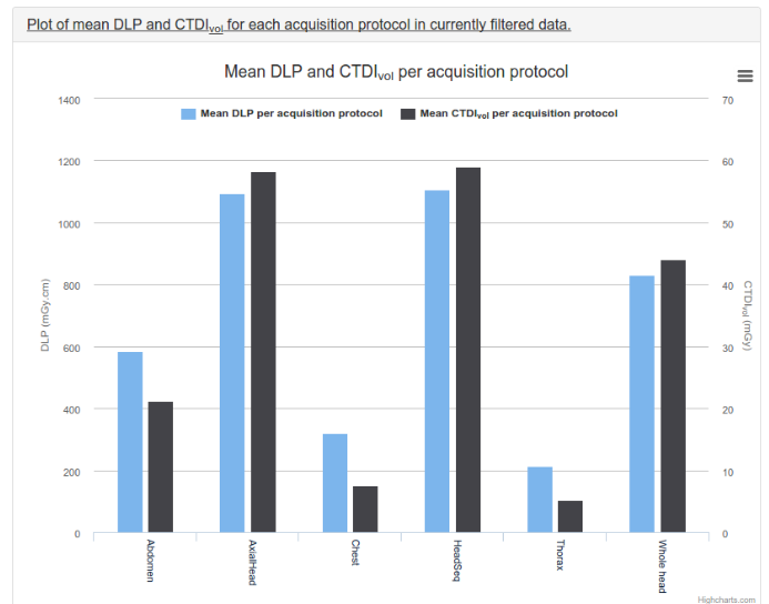
Charts and Visualization
Moreover, OpenREM offers visually intuitive charts for data exploration. These graphical representations provide an instant visual snapshot of exposure levels, thus facilitating insightful data analysis and informed decision-making.
Skin Dose Maps
A standout feature of OpenREM is its ability to generate skin dose maps for fluoroscopy. This feature allows for a detailed view of the radiation exposure across the patient's skin, thereby aiding in the identification of areas that may be at risk of excessive radiation exposure.
Export your Data
In addition to these features, OpenREM also boasts data export capabilities. This allows for the easy sharing and further analysis of data, enhancing the overall utility of the software.
Patient Privacy First
OpenREM prioritizes patient privacy. By default, patient identifiable data is not retained in the system. However, in situations where this data might be necessary, OpenREM can store it in a hashed format, conditioned upon the appropriate permissions being granted.
This ensures the security and confidentiality of sensitive patient information, reinforcing the trust between patients and healthcare providers.
Web Interface
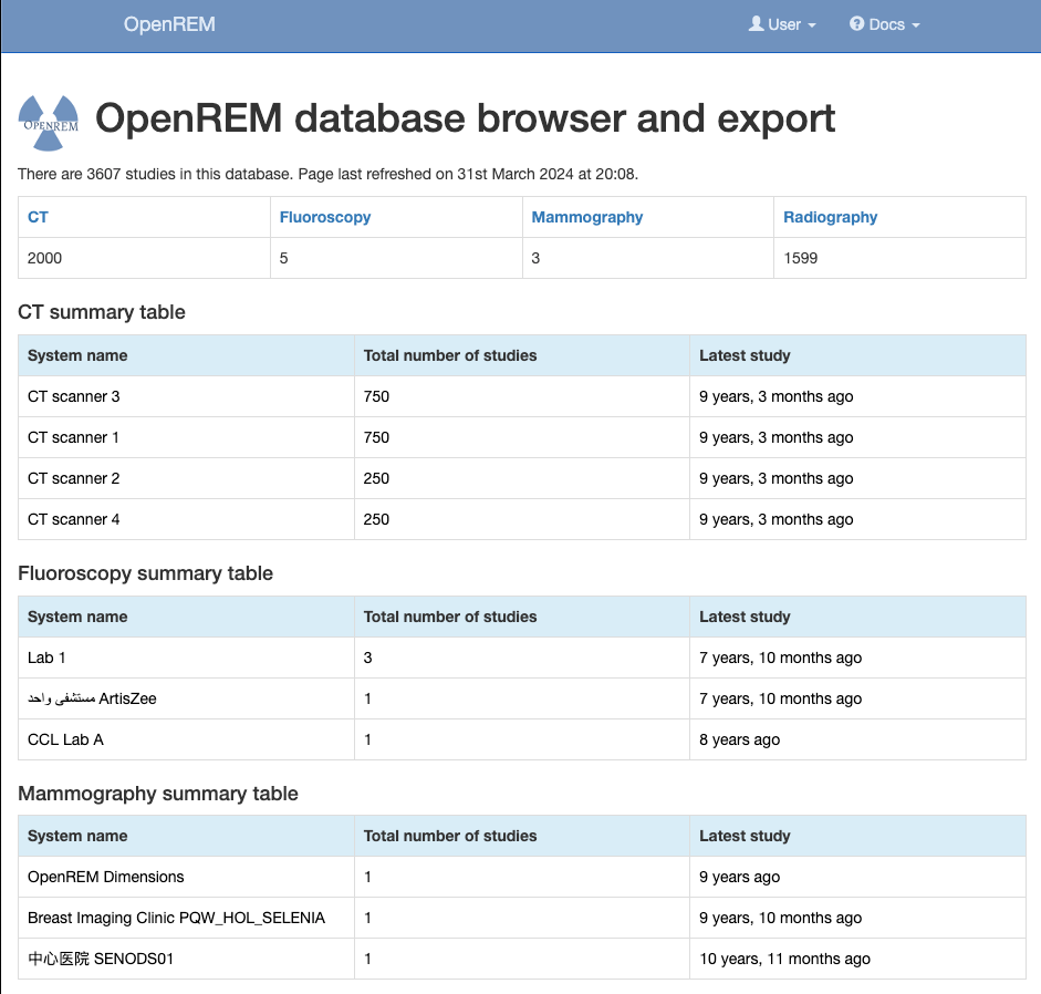
OpenREM provides a web interface for display of the studies that have been imported into the database, allowing easy review of the latest data. It also has a filtering function to enable any subset of the studies to be reviewed.
Each of the modalities has charts suitable for that modality optionally plotted within the web interface.
Features
Importing of CT, planar X-ray, fluoroscopy, and mammography data
Displaying summary exposure data, with filtering and searching
Charts to visualise and explore the data, including mean and median dose metrics, histograms, workload data and more
Skin dose maps for fluoroscopy using a simple geometric phantom
Export of data into spreadsheets, sorted and summarised
Patient privacy prioritization, with optional hashed storage of patient identifiable data
What is Niffler?
Niffler is a free open-source lightweight framework to facilitate executing machine learning pipelines and processing workflows on DICOM images and metadata. Niffler facilitates efficient transfer of DICOM images on-demand and real-time from PACS to the research environments.
Niffler is also integrated with the radiology information system (RIS)
With rising expenses and constant budget constraints in the healthcare sector, cost efficiency is becoming a top priority, and finding affordable yet effective solutions is more vital than ever. One such solution lies in the use of monochrome LCD screens. These screens reduce costs but maintain the high quality necessary
What is a portable DICOM viewer?
A portable DICOM viewer, is an app that you can run it directly to view DICOM files without installation.
It is a useful feature if you want to run, view and display DICOM images and media with portable media as USB-drive, DVD, or CD
Rubo Medical Imaging BV is a leading company that concentrates its efforts on the development of software based on the Digital Imaging and Communications in Medicine (DICOM) standards. This specialization allows them to provide a diverse range of tools that cater to different needs in the medical imaging field.
One
DWV (DICOM Web Viewer) represents a state-of-the-art open source zero footprint medical image viewer that stands out in the field of medical imaging.
The uniqueness of DWV lies in its exclusive use of javascript and HTML5 technologies, ensuring it is platform-independent and can seamlessly operate on any device that supports
The Dicom Image Viewer is an uncomplicated image view control tool that has been fully implemented in C#. This tool is especially useful when you need to export Dicom images.
As a medical image display control, it supports both dicom and raw image formats. This feature allows it to be
Blue Light is a sophisticated, browser-based medical image viewer that is primarily maintained and updated by the highly skilled team at the Imaging Informatics Labs. This platform is a pure single-page application (SPA), distinguished by its lightweight design.
It's exclusively developed using JavaScript and HTML5 technologies, which ensures
MRIcroGL is a cross-platform tool for viewing DICOM and NIfTI images, with a drag-and-drop interface and scripting language. It requires OpenGL 2.1 by default, but can also be compiled for OpenGL 3.3 Core or Metal for macOS users.
If OpenGL 2.1 is not supported, MRIcron can be
The Dicom Image Reader is an innovative, open-source medical image viewer, meticulously built using modern technologies such as JavaScript, HTML5, NodeJS, and the Electron framework.
This minimal yet powerful app is capable of loading and effectively manipulating data in DICOM format, a widely accepted standard in the medical imaging field.
Aliza MS is a free open-source DICOM viewer with a dozen of clinical-ready features.
It is available for Windows, Linux, and macOS. Because it is primarily written in C++ and C, It has a good performance even in working with large files and data sets.
Features
*
Very fast directory scanner,