BlueLight Viewer is a Free Web-based DICOM Viewer
Table of Content
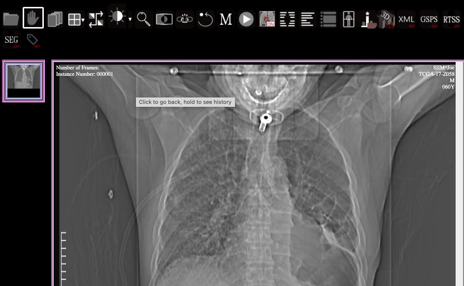
Blue Light is a sophisticated, browser-based medical image viewer that is primarily maintained and updated by the highly skilled team at the Imaging Informatics Labs. This platform is a pure single-page application (SPA), distinguished by its lightweight design.

It's exclusively developed using JavaScript and HTML5 technologies, which ensures its compatibility and easy deployment on any HTTP server. Just place it in the HTTP server, and it's ready to go.
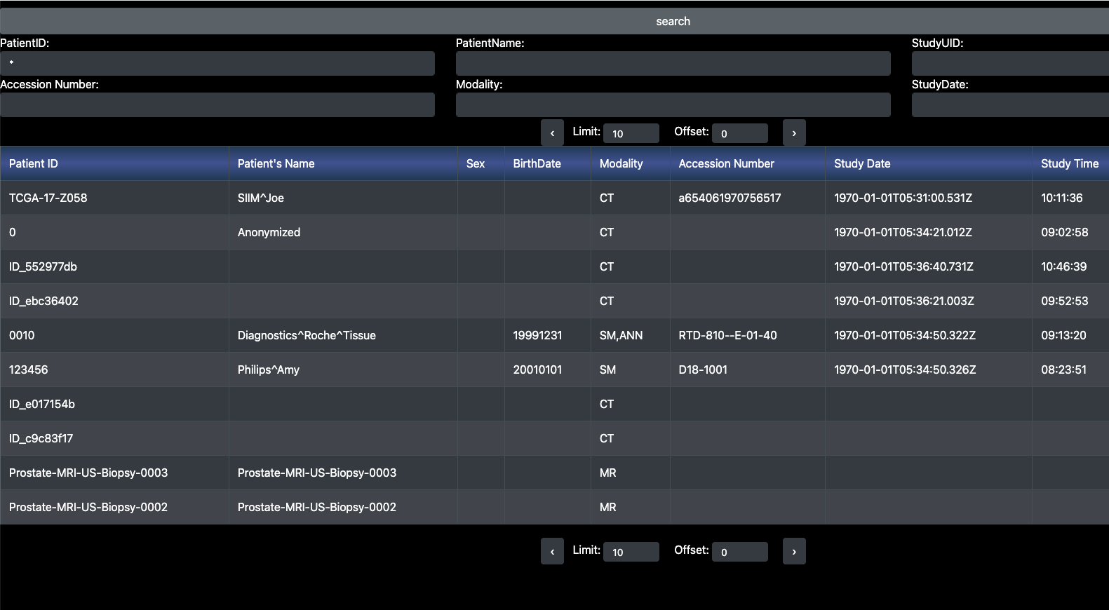
Beyond the convenience of its installation, Blue Light offers a wealth of functionality. It supports not just the opening and viewing of local data, but also the ability to connect to medical image archives that support the DICOMweb standard.
This means that it can access a vast range of medical imaging data stored remotely, making it an invaluable tool for healthcare providers.

Moreover, Blue Light is capable of displaying various image markups and annotations. These include Annotation and Image Markup (AIM), DICOM-RT structure set (RTSS), DICOM Overlay, and DICOM Presentation State. These are all critical tools for the interpretation and analysis of medical imaging data.
Adding to its impressive features, it provides tools specifically designed for medical image interpretation and 3D image reconstruction. Examples of these advanced tools include Multiplanar Reformations or Reconstructions (MPR) and Volume Rendering (VR). These tools further enhance the viewer's functionality, making it an indispensable tool in the healthcare industry.
Features
Network support
- Load local files
- Integration with any DICOMWeb Image Archive, including Raccoon, Orthanc, and dcm4chee server
- Retrieve methods: WADO-URI (as default) and WADO-RS: specify one of them in config.json
- Integration with XNAT by plugin xnat.js. BlueLight will query the XNAT's API to get the XML and retrieve the DICOM stored in experiments and its scans. Currently we doesn't build it as an XNAT plugin. issue: XNAT Connection
- Step1: copy BlueLight to XNAT deployment folder
- Step2: type URL: https://{XNAT's hostname}/bluelight/search/html/start.html?experiments={XNAT expID}&scans={scanID}&format=json
http://{XNAT's hostname}/REST/projects/test/subjects/XNAT_S00001/experiments/XNAT_E00002/scans/1/files?format=json
Support IODs
- Most general image IODs (CR, DX, CT, MR, US, etc)
Native features for 2D image interpretation
- Pan, zoom, move
- Scroll images within a series
- Rotation, Flip, Invert
- Windowing
- Cine
- viewports: 4×4
- Cross-Studies synchronization
- Magnifier, etc
- Line and angle measurement
- hide/display markups and annotations
- Export image
Support the display of the kinds of markups and annotations
- GSPS: DICOM Graphic Annotation
- DICOM Overlay
- DICOM-RT structure set (RTSS)
- Annotation and Image Markup (AIM)
- DICOM SEG (Segementation)
- LabelImg
- Provide the function to convert the DICOM Overalys to a DICOM SEG object.
Plugins Support
Bluelight separates advanced features into plugins for better performance. These plugins are located in the /scripts/plugin folder and can be enabled via the config. Examples include the 'oauth' and 'MPR' plugins.
New plugin ideas from third parties are welcome. Available plugins include Authentication, 3D Post-Processing, Labeling tool interfaces, and General plugins like Display DICOM TAG and Display LUT.
License
MIT License


